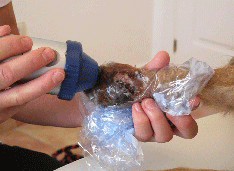
 ESWT in a Chow Chow with a non-healing wound
Four years prior to the first extracorporeal shockwave therapy session, the Chow Chow had injured her left metacarpal paw pad…
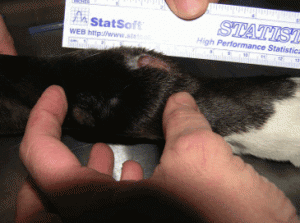 ESWT for Lick Granuloma in canines
8 year old Lab Mix breed dog treated with shockwave treatment for Lick Granuloma at his left carpal region.
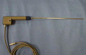 Urolith removal via a standing perineal urethrotomy in a quarter horse gelding using radial shock waves by William W. Ferguson, DVM, Rogue Valley Equine Hospital
_________________________________________________________________________________
ESWT Case Study – Cable Cut To The Hock
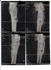 My most challenging and most successful result using shock wave therapy came from using the combined effects of the MASTERPULS MP50 shockwave machine and the IV laser on a mare who suffered a cable cut to her hock early in 2007. April 25, 2007 radiographs showed a sequestered bone 1-2 inches below the joint. Surgery was performed to remove the sequestered bone. Radiographs of 2007 showed the sequestered bone had combined in size and no further surgery. The mare suffered from extreme swelling and general inflammation of the surrounding tissue. I began treating this patient in early 2009 as a last chance effort to save her life. She was treated over separate treatment
 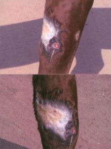 periods up to her final radiograph of October 2, 2009 which showed total resolution of the sequestered bone. The bone was intact with no evidence of the affected bone infection. Initially, the mare was treated for 30 days using ESWT, following with laser in between ESWT treatments. The combination of modalities brought the infection out through a visible lesion directly above the sequestered area shown by radiology. After 30 days, the leg swelling was reduced and the mare returned home. Two additional times she returned after her leg showed signs of swelling. She returned for a period of 7-9 days and received ESWT and laser treatment. Radiograph on October 9, 2009 showed the surprising results of combined treatment of ESWT and laser therapy.
Patricia Woodrick, Physical Therapist – Woodrick Ranch/ Light Therapy Partners – Aubrey, TX
READ ALSO PATRICIA WOODRICK’S TESTIMONIAL!
_________________________________________________________________________________
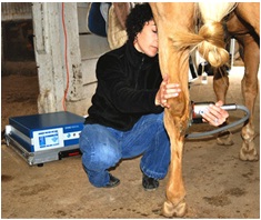 We use shock wave therapy for a number of applications. Suspensory desmopathy is one of the primary applications in our practice along with many other ligament and tendon injuries. We also treat non union fractures, dorsal spinous process disease (“kissing spines”) and certain osteoarthritis conditions.
CASE #1
One of our most interesting shock wave cases is a 12-year-old Westphalian gelding who was hit by a car around the first of the year. He was struck in such a way that, unknown at the time, the medial collateral ligament of his left hock was [EXPAND read more] torn along with significant bone trauma to the hock joints themselves. There was diffuse soft tissue damage and tearing of the attachments of the joint capsule. He had been rested and treated conservatively for 6 months as he surprisingly did not present initially with a significant lameness. It was not until several months later, when he got very excited and tore around his paddock that he came up acutely lame and his hock swelled severely. He then presented to us in early June for additional diagnostics and a further lameness workup. This horse was significantly lame and based on the diagnostic imaging results, his prognosis was guarded for a return to athleticism. We treated the medial collateral ligament and the dorsal and dorsomedial aspect of the hock (over the joint capsule) with the Storz Duolith focused probe with a medium energy level a total of 5 times on average two weeks apart. We also injected his distal hock joints and his tarsocrural joint and he has done remarkably well. This hock had been severely effused and had significant soft tissue swelling/thickening over the medial, dorsomedial and dorsal surfaces of the hock. The medial collateral ligament had significant fiber disruption and loss of integrity. We just saw this horse a few weeks ago, (approximately 4 months out from the start of treatment), for a lameness re evaluation. He is sound and the hock appearance is near normal. Ultrasonographically the collateral ligament is still larger than that of the contralateral limb but it has healed very well and has good fiber pattern and integrity. We have given the go ahead to gradually move forward from the light work he is currently in to a return to normal training. We can hardly believe he has recovered so well. It is truly a remarkable recovery.
CASE #2
Another recent case we treated with the STORZ DUOLITH VET is a 13-year-old Appendix Quarter Horse gelding that had been significantly lame in his left fore limb for approximately one week. He was referred in for a lameness work up and diagnostic imaging. This horse was a grade 3/5 lame on baseline. Nerve blocks followed by an ultrasonographic examination confirmed the lameness to be attributed to an injury to his left front deep digital flexor tendon (DDFT) at the level of the pastern. There was severe fiber disruption involving approximately 50 % of the deep flexor tendon. This horse received shock wave therapy over the damaged DDFT using a focused probe on a medium energy level (2000 impulses) at two week intervals. After three treatments this horse improved two full grades of lameness to a 1/5 LF baseline lameness. Prior to his fifth treatment (two months from the start of treatment) this horse was very nearly sound in a straight line and on the right circle and only a grade 1+ to 2- lame on the left circle. We have recommended the owner begin walking under saddle and that the horse return in four weeks for a follow up ultrasound exam to determine if structurally this horse can continue with a gradual return to previous levels of exercise. We are very encouraged by the progress seen so far and this horse’s response to treatment. [/EXPAND]
Patricia Quirion-Henrion, MA, NAVP – Equine Rehab Therapist
New England Equine Medical & Surgical Center, PLLC
www.newenglandequine.com
READ ALSO PATRICIA HENRION’S TESTIMONIAL!
_________________________________________________________________________________
Here are some great results we’ve gotten using the DUOLITH Shock Wave in our Equine Veterinary Practice:
- A 10-year-old Thoroughbred mare that was a Preliminary/Intermediate level event horse that went from too lame to ride on a front fetlock to competing at the previous level of competition after a series of 8 shockwave treatments and rest.
- A 20-year-old Hanoverian that was a former FEI level dressage schoolmaster still performing consistently at 4th level plagued by periodic bouts of severe cellulitis/lymphangitis that responds within 24 hrs of treatment with shockwave therapy when antibiotics and NSAIDs showed only partial response.
- A 6-month-old Thoroughbred filly with 4 deep non healing decubital ulcers healed quickly after shockwave therapy was initiated.
- An aged black & white Paint gelding with a non healing laceration and resulting proud flesh of 6 weeks duration. At time of admission to the hospital this gelding had a tennis ball sized wound. He healed without a scar.
- A 3-year-old Standardbred gelding with left carpal degenerative joint disease who was removed from competition and treated with rest and a series of shockwave treatments has since been returned to training and has raced successfully.
- A 10 year old warmblood eventing mare that was rejected on a pre-purchase exam for a stiff/sore back and a positive flexion test of a fetlock was treated with shockwave and is now performing at a higher level than at the time of the pre-purchase exame.
 Sarah Begley, DVM – Battenkill Veterinary Equine – Middle Falls, NY
_________________________________________________________________________________
For an additional selection of case studies or if you simply want to know more about FOCUS-IT or shockwave technology, please contact us.
FOCUS-IT, LLC
+1-770-612-8245